How peritoneal immune cells "remotely control" the healing of wounds
Immune cells in the abdomen can accelerate the healing of skin wounds, even in distant parts of the body. This has been shown by an international research team led by Inselspital, Bern University Hospital, and the University of Bern. The researchers developed a mouse model that they linked to patient data. The results offer new possibilities for predicting and treating wound healing disorders after surgery.
Wound healing disorders are among the most common and most serious complications, particularly after major abdominal surgery, in older people or in patients with diabetes. The human abdomen contains a large population of specialized immune cells that can significantly influence the healing process after injuries and operations. Understanding how the body controls tissue repair not only directly at the wound but also in the rest of the body is therefore of great medical and social importance.
A research team from the Department of Visceral Surgery and Medicine at Inselspital, Bern University Hospital and the Department for BioMedical Research (DBMR) at the University of Bern, in collaboration with the University of Calgary, has used mouse models and patient data to demonstrate that immune cells in the peritoneal cavity can influence wound healing via the bloodstream, even at distant sites in the body. The study highlights large macrophages in the abdominal cavity as key regulators of tissue repair. The results were recently published in the Journal of Clinical Investigation.
Innovative mouse model to study wound healing
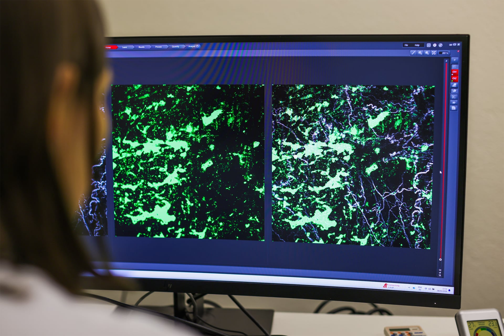
The starting point of the study was the observation that the peritoneal cavity - similar to the liver - contains large quantities of specialized immune cells. These macrophages are a type of immune cell that function like police officers in the body: they raise alarm when recognizing a potential threat. At the same time, they act as "vacuum cleaners" - hence also the name scavenger cells – by swallowing intruders such as bacteria or even damaged or old body cells. Until now, it was unclear whether these macrophages in the peritoneal cavity only support local healing processes or can also influence distant tissues such as the skin. To address this question, the team used a combined experimental and clinical research design: in mice, they created standardized skin wounds which were combined with targeted stimulation of the peritoneal cavity, and the blood, genes and key signaling molecules were analyzed in detail using state-of-the-art methods.
In parallel, the researchers examined blood serum samples and follow-up data from patients who had undergone major abdominal surgery at the Inselspital. "We developed a model that allows us to directly link local abdominal surgery and the healing of remote skin wounds," says first author of the study Dr. Lilian Salm, senior physician at the Department of Visceral Surgery and Medicine at Inselspital, Bern University Hospital and researcher at the Department for BioMedical Research. She adds: "The combination of mouse models, high-resolution molecular and microscopic analyses and clinical patient data makes our results particularly robust and directly relevant for surgery."
Immune cells in the abdomen act similarly to hormones
The experiments showed that stimulation of the peritoneal cavity – either as part of a surgical procedure of the abdominal cavity or through inflammatory stimuli – significantly accelerates the healing of remote skin wounds. Using imaging techniques and the newly developed mouse model the researchers were able to show that the large peritoneal macrophages do not migrate to the skin wound themselves. "Instead, once activated, they release the protein fibronectin into the bloodstream. This protein then accumulates specifically in the wound area and promotes wound healing there," explains Prof. Dr. Guido Beldi from the Department of Visceral Surgery and Medicine at Inselspital, Bern University Hospital and research group leader at the Department for BioMedical Research at the University of Bern. He adds: "If these macrophages are selectively removed from the peritoneal cavity in mice or the fibronectin gene is switched off, the positive effect on wound healing disappears completely; the administration of fibronectin, on the other hand, can restore it." These findings help explain why wound healing is impaired in some patients after surgery: "Thanks to the experimental data and, in particular, the combination with clinical data, we now understand why wound healing does not function optimally in some patients. In these individuals, we observed low plasma fibronectin levels, indicating insufficient activation of macrophages.”
This represents a paradigm shift for research: plasma fibronectin - i.e. fibronectin that dissolves in the blood - originates not only from the liver, but also to a relevant extent from macrophages in the peritoneal cavity. The results expand our understanding of macrophages, which not only act as local "cleaning and repair cells", but also act as regulators like hormones and influence distant tissue via the bloodstream. "Our work shows that the peritoneal cavity is a previously underestimated hormone-like control center for wound healing," says Beldi. "This opens up new perspectives for the systemic investigation of phagocyte-dependent signaling pathways not only in injuries, but also in tumor biology, blood poisoning or aging processes."
New opportunities for the prediction and treatment of wound healing disorders
The study points beyond basic research to specific clinical applications. Fibronectin could be suitable as a biomarker to better assess the individual risk of wound healing disorders after major surgical procedures. Early identification of high-risk patients in particular would be of great benefit. "Our next step is to investigate whether fibronectin serum levels can be used to predict wound healing disorders after major abdominal surgery," says Salm. "In parallel, we will investigate whether a targeted increase in fibronectin or the activation of macrophages in the peritoneal cavity can improve recovery after surgery in high-risk groups." The University of Bern and Inselspital, Bern University Hospital have internationally renowned expertise in the field of immune defense in the abdominal cavity. "Our research group combines clinical visceral surgery with experimental immunology of the peritoneal cavity in a unique way," emphasizes Beldi. "Direct access to surgical patient data, state-of-the-art imaging such as intravital microscopy and close collaboration with partners such as the University of Calgary enable us to quickly translate basic mechanisms of the immune response into clinically relevant concepts," concludes Beldi.
Publication details:Salm L, Zwicky S, Spari D (2026) Peritoneal macrophages regulate distal wound healing via endocrine release of plasma fibronectin. Journal of Clinical Investigation. Published online on May 1, 2026. |
Department for BioMedical Research (DBMR)The Department for BioMedical Research (DBMR) of the Faculty of Medicine of the University of Bern was founded in 1994 by the University of Bern and the Inselspital, University Hospital Bern. The DBMR is divided into 13 research programmes with around 100 participating individual laboratories and several independent research laboratories whose research spans all biomedical areas. To bridge the gap between the laboratory and the bedside, the DBMR promotes clinical research with a strong emphasis on the development of translational approaches, the use of "omics" and other cutting-edge technologies, and extensive collaboration between laboratory-based and patient-centered clinical research. The DBMR is also committed to the promotion of young scientists. |
Visceral Surgery and Medicine, Inselspital, Bern University HospitalFurther information: https://bauchzentrum-bern.ch/de/ |
05.05.2026