Controlling adhesions in the abdomen
Adhesions are scars in the abdomen, which can occur after surgery, often have serious consequences. Now, researchers from the University of Bern and Inselspital, University Hospital Bern, in collaboration with Canadian researchers, have discovered how such adhesions form. The findings may help to develop a drug to prevent adhesions in the future. The study was published as the cover story of Science magazine.
Scars inside the abdomen, known as adhesions, form after inflammation or surgery. They can cause chronic pain and digestive problems, lead to infertility in women, or even have potentially life-threatening consequences such as intestinal obstruction. If adhesions develop, they must be operated on again. They also make subsequent surgical interventions more difficult. This leads to substantial suffering for those affected and is also a significant financial burden for the healthcare system. In the USA alone, adhesions in the abdomen result in healthcare costs of 2.3 billion dollars per year.
Knowledge about the cause of adhesions is still incomplete, and there no therapy for them. "Because the disease has been largely overlooked in research, we have started this program in Bern to find out more about the development of adhesions," says Daniel Candinas, Co-author of this study. It had already been suspected that special immune cells, called macrophages, play a decisive role in the development. This was confirmed by Joel Zindel and Daniel Candinas from the Department of Visceral Surgery and Medicine at Inselspital and Department for BioMedical Research (DBMR) at the University of Bern.
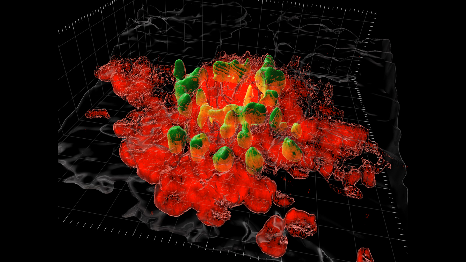
Then, Zindel continued his research at the University of Calgary in Canada in the group led by Paul Kubes, as they are considered world-leading in the field of macrophages in the abdominal cavity. Thanks to Zindel's clinical expertise and the Canadian researchers’ know-how, it was possible to develop a new imaging system using cutting-edge microscopy that allows to see inside the living body. This allowed them to catch the macrophages in flagrante and on film, as they form shapes that then lead to the adhesions.
The researchers were also able to describe the molecular mechanisms behind this. The results of the study have now been published as the cover story of the journal Science.
New technology developed
Macrophages are found in what is called peritoneal fluid, a lubricant between the peritoneum, which is inner lining of the abdominal wall, and a similar lining around the organs in the abdominal cavity. Macrophages passively swim around in this fluid, much like plankton in the sea. Their tasks include eliminating pathogens, but also sealing injuries in the abdominal cavity as quickly as possible.
How they accomplish the latter, i.e., recognizing an injury and moving there, was unclear until now. Since these cells behave in the test tube very differently from the way they do in the body, Zindel and Kubes developed a new microscopy technique that allowed them to use the thinnest part of the abdominal wall as a window to look into the peritoneal caity, the “native habitat” of these macrophages and film them as they move arround.
When macrophages lose control
When there is an injury within the abdominal cavity, macrophages aggregate within minutes to form clot-like structures. In this way, they seal the injury. As the researchers led by Zindel and Kubes have discovered, the molecular mechanism behind this is based on special, non-specific receptors that recognize a variety of structures. Simply by being moved through the fluid via respiratory or digestive movement, these receptors act to initiate clotting around a wound. What works fine for smaller injuries becomes a problem for large ones, however, such as surgically opening the abdominal wall, or inserting an implant. "In larger injuries, the macrophages get out of control - the clots don't stop growing and form long strands," Zindel explains. "We were able to show that these strands are what lead to the adhesions."
This could have evolutionary reasons: Macrophages are optimized by evolution to cope well with small injuries. "Let's take the example of a hunter who is injured by a deer antler," Zindel says. "The macrophages seal all the internal holes as quickly as possible – which is the only way to survive." However, when air enters the abdominal cavity during abdominal surgery or foreign bodies are implanted, the macrophages are overwhelmed because evolution has not prepared them for this challenge. "In this case, the macrophages become harmful and cause adhesions," Zindel explains.
Patent pending
The researchers found that when the corresponding receptors are blocked in mice, it leads to fewer adhesions. Thus, a patent application has been filed for the use of the active ingredient against adhesions. The findings are relevant to many areas of research, as there are no other immune cells which are being recruited as quickly as macrophages in the abdominal cavity. Similar mechanisms could be present in other cavities such as the heart or lungs, or could play a role not only in injuries but also diseases such as abdominal tumors.
"This is an example of how laboratory research has high translational value," says Daniel Candinas. Together with Calgary, the Bernese researchers will now look for partners in the industry and are hopeful that they will be able to confirm the efficacy of the compound in human tissue as well. In the future, patients could, for example, be given a drug before surgery that suppresses the macrophage reaction and inhibits the formation of adhesions.
Publication details:J. Zindel, M. Peiseler, M. Hossain, C. Deppermann, W.Y. Lee, B. Haenni, B. Zuber, J.F. Deniset, B.G.J. Surewaard, D. Candinas, P. Kubes: Primordial GATA6 macrophages function as extravascular platelets in sterile injury, Science, 4 March 2021,https://science.sciencemag.org/content/371/6533/eabe0595 |
Department for Biomedical Research (DBMR)The Department for BioMedical Research (DBMR) of the Faculty of Medicine of the University of Bern, led by Prof. Dr. med. Mark Rubin, was established in 1994 by the University of Bern and the Inselspital (Bern University Hospital). To realize its mission to bridge the gap between bench and bedside, the DBMR promotes an integrative perspective to clinical research with a strong emphasis in the development of translational approaches, the use of omics and other cutting-edge technologies, and extensive interaction and collaboration between laboratory-based and patient- oriented clinical research. The DBMR is also committed to fostering the careers of young academics. |
2021/03/05


